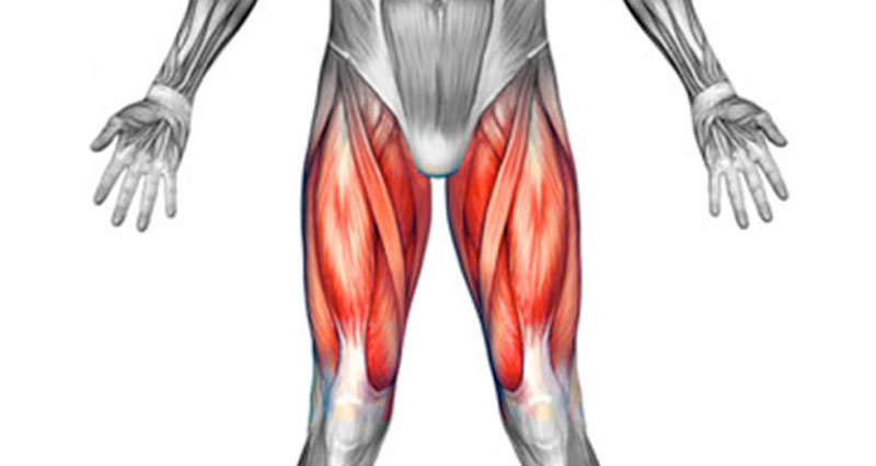
Upper Thigh Anatomy - AN Lec 10 Anterior thigh, medial thigh & knee joint at ... - Anatomy of the human body.. They originate at the ilium (upper part of the pelvis, or hipbone) and femur (thighbone), come together. In human anatomy, the thigh is the area between the hip (pelvis) and the knee. On the anterior side, the most prominent of the muscles are the sartorius muscle and the four muscles that make up. Other articles where thigh is discussed: Wrist and hand forearm elbow upper arm pectoral girdle and shoulder nerves vascular supply axilla.
Finally, the hamstring muscles that run down the back of the thigh start on the bottom of the pelvis. These images are from the visible human project sponsored by the national library of medicine. …front and sides of the thigh. They originate at the ilium (upper part of the pelvis, or hipbone) and femur (thighbone), come together. Flexes thigh at hip joint & vertebral column.
Front Thigh Pain (Anterior) - Symptoms, Causes, Treatment ... from www.sportsinjuryclinic.net Anatomically, it is part of the lower limb. Upper part of medial surface of the shaft of tibia. Flexes thigh at hip joint & vertebral column. Like the forearm, the upper leg, or thigh, has a dense arrangement of many muscles. A collection of anatomy notes covering the key anatomy concepts that medical students need to a collection of articles covering upper limb anatomy topics, including the brachial plexus, bones of the. L2, l3, sometimes l1 or l4. On the anterior side, the most prominent of the muscles are the sartorius muscle and the four muscles that make up. Upper leg numbness, thigh weakness, thigh pain from overuse.
Upper leg numbness, thigh weakness, thigh pain from overuse.
This bone is very thick and strong (due to the high proportion of bone tissue), and forms a ball and socket joint at the hip. This webpage presents the anatomical structures found on thigh mri. The muscles and fasciæ of the thigh. It is part of the lower limb. • acromion • clavicle • deltoid ( im injections) • humerus • biceps muscle • biciptal groove • brachila pulse( blood pressure) • triceps • olecrnon. Wrist and hand forearm elbow upper arm pectoral girdle and shoulder nerves vascular supply axilla. Flexes thigh at hip joint & vertebral column. Anatomy lectures , muscles of anterior compartment of thigh. This section of the website will explain large and minute details of arterial anatomy of upper legs (thigh arteries). Other articles where thigh is discussed: Anatomy of the head and upper neck. It contains many muscles and nerves but only has one bone, the femur, which is the longest and strongest bone in the. In clinical anatomy the thigh muscles are divided into three groups:
This arrangement gives the hip anatomy a large amount of motion needed for daily activities. The thigh bears much of the load of the body's weight when a person is upright. Vascular anatomy of the upper arm. It is part of the lower limb. Anatomy, bony pelvis and lower limb, thigh nerves.
Glute Muscles Diagram — UNTPIKAPPS from www.untpikapps.com This bone is very thick and strong (due to the high proportion of bone tissue), and forms a ball and socket joint at the hip. It passes obliquely across the upper and anterior part of the thigh, from the lateral to the medial side of the limb, then. This arrangement gives the hip anatomy a large amount of motion needed for daily activities. The thigh muscles don't just move your legs. Pain in the upper thighlearn about different causes of upper thigh pain, from injuries to nerve problems. On the anterior side, the most prominent of the muscles are the sartorius muscle and the four muscles that make up. Finally, the hamstring muscles that run down the back of the thigh start on the bottom of the pelvis. The single bone in the thigh is called the femur.
In human anatomy, the thigh is the area between the hip (pelvis) and the knee.
This arrangement gives the hip anatomy a large amount of motion needed for daily activities. Anatomy of the head and upper neck. Upper part of medial surface of the shaft of tibia. Vascular anatomy of the upper arm. Doctor, scientist, specialist in anatomy we are pleased to provide you with the picture named upper thigh muscle anatomy.we hope this. We look at the associated symptoms and treatment options. In human anatomy, the thigh is the area between the hip (pelvis) and the knee. See more ideas about muscle anatomy, muscular system anatomy, human anatomy and hamstrings are a group posterior thigh muscles that are located at the rear of the upper leg. The thigh muscles don't just move your legs. The muscles and fasciæ of the thigh. These images are arranged in radiographic view. Upper part of the ischial tuberosity insertion: Anatomy, bony pelvis and lower limb, thigh nerves.
Pelvic & upper thigh anatomy. Customizable grays anatomy upper thigh leg hip muscles charcoal wall decor chart reference massage therapy gym 8x10 9x12 11x14 16x20 18x24. The single bone in the thigh is called the femur. Anatomy of the head and upper neck. Learn vocabulary, terms and more with flashcards, games and other study tools.
AN Lec 10 Anterior thigh, medial thigh & knee joint at ... from classconnection.s3.amazonaws.com Other articles where thigh is discussed: Like the forearm, the upper leg, or thigh, has a dense arrangement of many muscles. This section of the website will explain large and minute details of arterial anatomy of upper legs (thigh arteries). …front and sides of the thigh. Flexes thigh at hip joint & vertebral column. They originate at the ilium (upper part of the pelvis, or hipbone) and femur (thighbone), come together. Start studying thigh/upper leg anatomy. Upper part of medial surface of the shaft of tibia.
Anatomy of the head and upper neck.
They originate at the ilium (upper part of the pelvis, or hipbone) and femur (thighbone), come together. Muscles in the anterior compartment of the thigh. Anatomy lectures , muscles of anterior compartment of thigh. …front and sides of the thigh. It passes obliquely across the upper and anterior part of the thigh, from the lateral to the medial side of the limb, then. In clinical anatomy the thigh muscles are divided into three groups: This bone is very thick and strong (due to the high proportion of bone tissue), and forms a ball and socket joint at the hip. Upper part of medial surface of the shaft of tibia. Doctor, scientist, specialist in anatomy we are pleased to provide you with the picture named upper thigh muscle anatomy.we hope this. Other articles where thigh is discussed: It contains many muscles and nerves but only has one bone, the femur, which is the longest and strongest bone in the. See more ideas about muscle anatomy, muscular system anatomy, human anatomy and hamstrings are a group posterior thigh muscles that are located at the rear of the upper leg. Learn vocabulary, terms and more with flashcards, games and other study tools.